Below I will share with you some diagnostic methods for skin diseases in dogs and cats and the key points for diagnosis of ten common skin diseases. Hope it helps you!
Diagnosis method
11. Consultation
(I) Disease course
The manifestations of animals in the early stages of the disease; what medicines have been used, whether the symptoms gradually decrease or continue to aggravate after taking the medicine; whether the living environment of dogs and cats, whether they often go to the grass to play; whether they have ever been exposed to sick dogs and cats; what shampoo is used, how to use shampoo, and how to take a bath; which part of the dog and cat skin is ill, whether it is itchy, etc.
(II) Medical history
Have you suffered from mite infection or fungal infection; whether you are in the late stage of delivery; whether you have a history of drug allergy, a history of contact dermatitis and a history of infectious diseases.
2. Generally, check
(I) Local skin observation: whether the hair is reversed, whether it has luster, whether it has hair loss, whether it is bilateral, local skin elasticity, stretchability, thickness, pigmentation, etc.
(Bi) Lesion situation
location, size, shape, concentrated or scattered, unilateral or symmetrical, surface conditions (bulge, flat, sunken, moist, etc.), smooth or rough, wet or dry, hard or soft, large or small elastic, local color, etc.
3. Laboratory inspection
(I) Parasite inspection
1. Cellophane tape inspection: that is, stick transparent tape with your hands, reverse hair sampling, and parasites are easily found.
2. Skin material examination.
3. Fecal examination.
(II) Fungal examination
1. Microscopic examination: The hair should be wider. After wrinkling the skin, scrape it to the dermis with a blade. After bleeding, put the scraping and extract on the slide.
2. Wood’s lamp examination
3. Fungal culture: (add US Moore indicator) Hair is taken at the junction of the health site and the lesion.
(III) Bacterial examination
Direct smear or contact specimens are subjected to staining, bacterial culture and drug sensitivity tests, etc.
(IV) Skin allergy test
After local scissors and shaving, use a syringe equipped with skin allergic reagents to perform different allergen tests in points. If yellow papules appear locally, they are allergic.
(V) Pathological histological examination
Direct smear or biopsy.
(VI) Allergic reactions
Intradermal reactions and patch tests.
(VII) Immunological examination
Immunofluorescence examination.
(VIII) Secretion function checks the functions of thyroid, adrenal and gonads.
The key points for diagnosis of common skin diseases
1. Flea infection
1. It is mainly pruritus scratching and rash on the back and neck;
2. It is common in summer and autumn;
3. It is wandering in the itching area.
2. Tick disease
Ticks are found to be parasitic on dogs. In most cases, dog owners see and peel off the colored "worms" on the dog's body when grooming the dog's hair. This is the tick, which ranges from millet grains to soybeans.
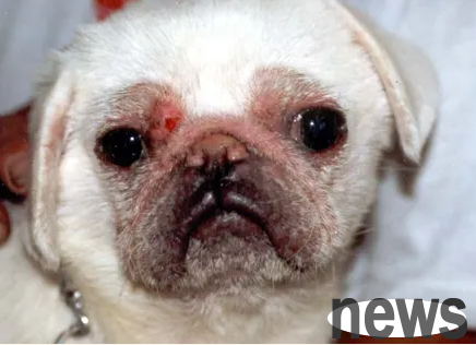
3. Canine Demodex infection
Dog Demodex parasites in the hair follicles of the dog's skin, and mostly parasitizes in the blister-like protrusions of the skin, and completes the life history here, which takes 24 days in total. A wide number of erythemas with distinct boundaries from the surrounding area can be seen on the skin. Red spots often appear in hairless areas on the eyes, ears, lips and inner legs, and the dog does not feel itchy.
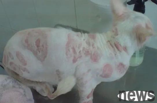
Disdemodex disease is a parasitic disease that can cause death of dogs. It has severely infected dogs, with large-scale hair loss and swelling. When erythema, seborrhea and purulent dermatitis occur, the diseased dogs are itchy and have common surface lymph node lesions.
4. Scabia mite infection
Dogs generally have symptoms such as hair loss, thickening of skin, erythema, small pieces of scab and scales, and very itching, causing the dog to scratch itself, secondary bacterial infection. Scabies often parasitize in the external ear, and in severe cases it affects the elbow and tarsal joints. Clinically, there are many common lower abdomen lesions.
5. Ear mite infection
Early infection of ear itchy mites in dogs and cats is often bilateral. Further development will result in widespread infection of the entire auricle, with obvious scales, excessive keratosis and scratching themselves. More severe infections include thick hyperkeratotic scales on both auricle and spread to the front of the head. Ear mites in dogs and cats often invade the external auditory canal, but ear mites can also cause itchy dermatitis in the ear and tail tips.
6. The infection of syllium mites by syllium mites causes mild itching, and yellow-gray scales on the dog's back, buttocks, head and nose, falls off during exercise. Some dogs also have insects but are asymptomatic. Jizhemi can infect people.
7. Lice disease
(I) Hairy lice
Because dog hair lice feeds on hair and epidermal scales, it causes itching and restlessness to the dog. The dog bites the itchy area and causes self-damage, causing hair loss, secondary eczema, papules, blisters, pus bubbles, etc. In severe cases, poor appetite will affect the dog's sleep and cause malnutrition in the dog.
(II) Long-jaw lice
Dog long-jaw lice are blood-sucking parasites. When sucking blood, it secretes toxic liquid, which stimulates the nerve endings of the dog and creates an itchy feeling. A large number of infections can cause suppurative dermatitis, which can cause hair loss or hair loss. The dog is depressed and weak, and has anemia due to chronic blood loss. The dog has poor resistance to other diseases.
8. Hookworm larval dermatitis
bend nematode, parasitizing in the small intestine of dogs, mainly in the duodenum, and also infects foxes and cats.
The main symptoms are weight loss, pale conjunctiva, easy hair loss, vomiting, diarrhea alternating between diarrhea, bloody or black feces. Infectious larvae invade the dog's claws (between fingers and toes), causing chronic itching dermatitis, and pustules may appear.
9. Microfilariatic dermatitis of canine fermentia
The canine fermentia fermentia
The canine fermentia
The intermediate hosts are fleas and mosquitoes. The main symptoms are: dyspnea, anemia, and circulation disorders; at the beginning of the disease, the dog has chronic cough, erythema of the skin, and sometimes itchy multiple focal nodules (suppurative granulomas in the blood vessels)
Diagnosis: Skin scraping (blood seepage) or tissue biopsy, it was found that the microfilariatic nature of the canine oxidia (in the subcutaneous capillaries)
10, tinea disease
10, tinea disease
10, tinea disease is a disease caused by fungal infection of the skin, hair and nails.
Canis tinea disease is mainly caused by Microsporidium canis, followed by Microsporidium gypsum-like and Hairy Beard-beard-beard-beard-beard-beard-beard-beard-beard-beard-beard-beard-beard-beard-beard-beard-beard-beard-beard-beard-beard-beard-beard-beard-beard-beard-beard-beard-beard-beard-beard-beard-beard-beard-beard-beard-beard-beard-beard-beard-beard-beard-beard-beard-beard-beard-beard-beard-beard-beard-beard-beard-beard-beard-beard-beard-beard-beard-beard-beard-beard-beard-beard-beard-beard-beard-beard-beard-beard-beard-beard-